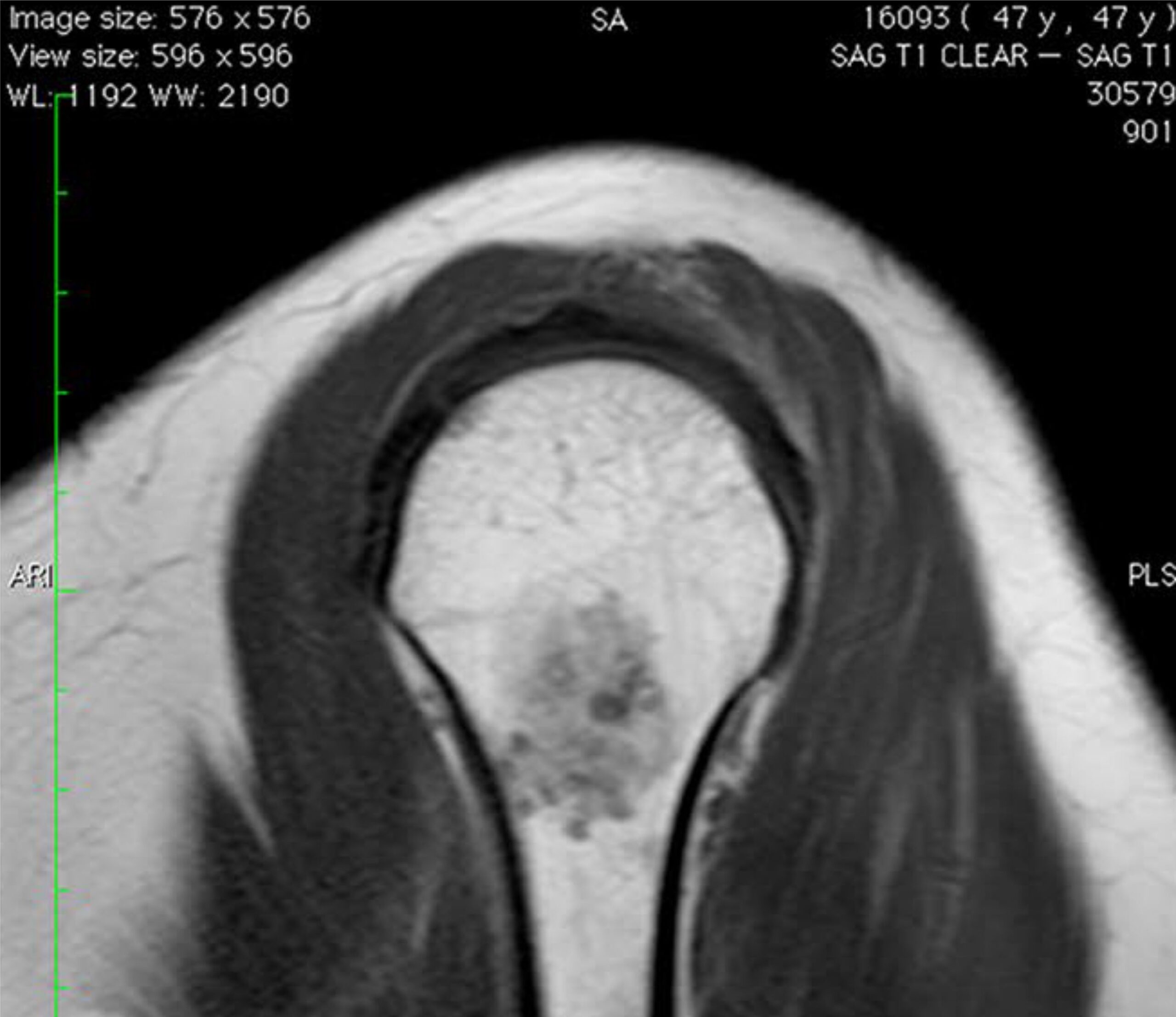
The differential diagnosis between chondroma and intraosseous chondrosarcoma is based on imaging and clinical exams, but only a biopsy can confirm diagnosis. The aim of this study was to evaluate the value of PET–CT in differen‑ tially diagnosing chondroma and chondrosarcoma. From October 2009 to May 2015, 36 patients with cartilaginous bone lesions in the extremities, 12 (33.3 %) men and 24 (66.6 %) women, were prospectively included in the study. Patients ranged in age from 21 to 68 years, with a mean age of 44 years. Lesions were located in the long bones: in the proximal humerus in 26 (72.2 %) patients, in the femoral shaft in 1 (2.7 %), in the distal femur in 7 (19.4 %), and in the proximal tibia in 2 (5.5 %). The SUVmax value of 2.0 was used to separate between patients submitted to surgery and patients submitted to observation. Among the 36 patients studied, 17 (47.2 %) had SUVmax ≤ 2.0, and they were diagnosed as chondroma and they were treated conservatively. Follow-up ranged from 14 to 76 months, averag‑ ing 38 months. Nineteen (52.7 %) patients with SUVmax >2.0 were diagnosed as chondrosarcoma and underwent surgery. The area of the curve, calculated considering the SUV variable as numeric, is estimated in 0.966, with a 95 % confidence interval from 0.906 to 1.000. To evaluate the sensitivity, specificity and positive/negative predictive values, it was built a 2 × 2 table. Significance was set at p < 0.05. According the criteria of maximum sensitivity and specific‑ ity, the cut point suggested to SUVmax was 2.2. If we consider this point, it is possible to identify 19 of 36 positive cases to chondroma (52.8 %), it means, all chondrosarcomas of the series. We concluded that PET–CT can be used as an objective and quantitative method of differentiating between chondromas and chondrosarcomas located within the long bones. It represents a complementary examination to standard imaging (X-ray, scintigraphy, CT and MRI) and pathological exams. The SUVmax between 2.0 and 2.2 would be a range area between chondroma and chondrosar‑coma and this range can be of value, among others exams, in decide the best treatment for patients with cartilagi‑nous lesions in long bones.
Baixe o artigo completo clicando aqui!